Coenzyme Q10


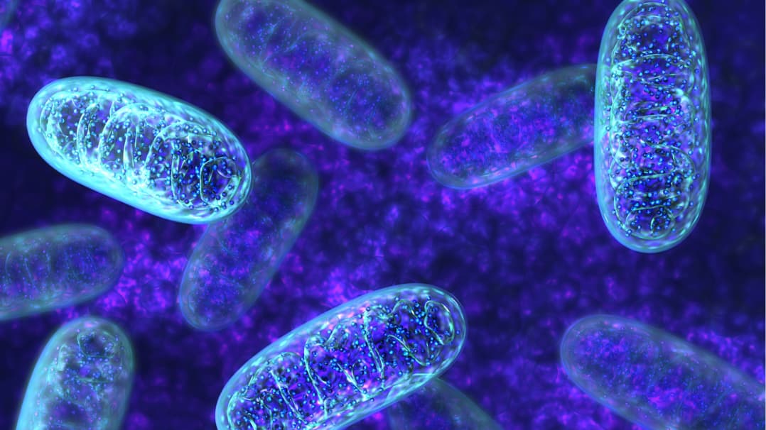
Coenzyme Q10 (CoQ10) is an important fat-soluble nutrient, because it’s essential for cellular energy production (i.e., ATP) and antioxidant defenses, helping protect membranes from oxidative stress. Because of its central role in ATP generation, the highest amounts of CoQ10 are found in organs that use the most energy, like the heart, liver, and kidneys. Meat and fish, especially their organs, are very good food sources. The best vegetarian sources are foods high in fat, including nuts, seeds, avocados, and vegetable oils. A person eating an average diet will get about 3-6 mg of CoQ10 a day [1–4]. Most CoQ10 isn't from diet, it's made in the body (i.e., biosynthesized), with creation requiring at least 12 genes. While the human body can make CoQ10, it may not always be able to make enough to meet its needs [5]. This seems to occur with aging, because CoQ10 gradually declines with age in a number of different tissues [6,7].*
Top Benefits of CoQ10
Supports mitochondrial structure and function*
Supports antioxidant defenses*
Support healthy cardiovascular function*
Supports healthy aging*
Qualia’s CoQ10 Sourcing
CoQ10 is most commonly supplemented in its oxidized form, which is called ubiquinone. Most human clinical studies have been done with the ubiquinone form. It can also be supplemented in its reduced ubiquinol form.
CoQ10 sourcing is focused on ensuring it is non-GMO, gluten-free and vegan.
CoQ10 Formulating Principles and Rationale
CoQ10 is dose-dependent (see Qualia Dosing Principles) in the range it’s commonly dosed (30 mg to several hundred milligrams a day). Body stores are maintained by a combination of the CoQ10 we consume in foods and supplements, and the CoQ10 made in our body [7]. It’s been suggested that a daily intake ranging from 30–100 mg in otherwise healthy persons is a good range to maintain healthy levels [7,8]. CoQ10 is additive with other mitochondrial and antioxidant nutrients. This means lower amounts of CoQ10 are often needed to support healthy function when it is combined with other nutrients, compared to when it is given as an isolated nutrient.*
CoQ10 Key Mechanisms
Supports mitochondrial structure and function*
CoQ10 is part of the electron transport chain of the inner mitochondrial membrane [9]
CoQ10 transfers electrons from complexes I and II to complex III by undergoing redox cycles between its three redox states (ubiquinone [fully oxidized], ubisemiquinone, and ubiquinol [fully reduced])* [9]
CoQ10 is critical in ATP generation via the electron transport chain* [9]
Supports electron transport chain performance and ATP production* [10–13]
Supports the NAD+ pool (NAD+/NADH ratio)* [10]
Supports β-oxidation* [14]
Supports mitochondrial membrane potential* [13,15]
Supports transcription factors of mitochondrial biogenesis (PGC-1α, NRF2, TFAM)* [10,16]
Supports mitochondrial DNA (mtDNA)* [10]
Supports mitochondria mass* [10,13]
Supports healthy lysosomal function*
Supports the transport of protons across lysosomal membranes to maintain the optimal pH* [9]
Supports the activity of digestive enzymes within lysosomes* [9]
Supports the lysosomal digestion of cellular debris* [9]
Supports antioxidant defenses*
Coenzyme Q10 (as ubiquinol) is a potent lipid-soluble antioxidant* [9,17,18]
Supports antioxidant defenses* [11,12,19]
Counters ROS production and oxidative stress* [7,10,15,17,20,21]
Replenishes glutathione (GSH) levels* [10]
Supports the regeneration of the lipophilic antioxidant alpha-tocopherol (vitamin E)* [9,18]
Promotes a healthy metabolic function*
Supports healthy lipid metabolism* [16]
Influences peroxisome proliferator-activated receptor gamma (PPARγ) activity* [16]
Supports UCP1 activity and the thermogenic function of brown adipose tissue* [16]
Supports healthy cardiovascular function*
Supports healthy cardiac function* [22–24]
Supports healthy endothelial cell oxidative stress responses* [25,26]
Supports endothelial progenitor cells* [15]
Supports endothelial/vascular function and blood flow* [27–30]
Promotes healthy aging and longevity*
Supports neuroprotective functions* [21,31]
Supports SIRT1 and SIRT3* [10,16]
Supports AMPK activity* [10,14–16,25]
Supports PPARα activity* [10,14,16]
Supports LKB1 activity* [10]
Supports DNA structure* [32]
Complementary ingredients*
Lipoic acid — support of mitochondrial function* [33–36]
Creatine — support of neuroprotection and of mitochondrial function* [33,34,37]
L-carnitine — support of mitochondrial function* [38]
Piperine — support of CoQ10 bioavailability* [39]
Vitamin B3 (NAD+ precursors) — support of mitochondrial performance* [40]
Vitamin E — support of mitochondrial function* [35]
*These statements have not been evaluated by the Food and Drug Administration. This product is not intended to diagnose, treat, cure, or prevent any disease.
REFERENCES
[1]C. Weber, A. Bysted, G. Hłlmer, Int. J. Vitam. Nutr. Res. 67 (1997) 123–129.
[2]P. Mattila, J. Kumpulainen, J. Food Compost. Anal. 14 (2001) 409–417.
[3]H. Kubo, K. Fujii, T. Kawabe, S. Matsumoto, H. Kishida, K. Hosoe, J. Food Compost. Anal. 21 (2008) 199–210.
[4]I. Pravst, K. Zmitek, J. Zmitek, Crit. Rev. Food Sci. Nutr. 50 (2010) 269–280.
[5]J.D. Hernández-Camacho, M. Bernier, G. López-Lluch, P. Navas, Front. Physiol. 9 (2018) 44.
[6]A. Kalén, E.L. Appelkvist, G. Dallner, Lipids 24 (1989) 579–584.
[7]L. Ernster, G. Dallner, Biochim. Biophys. Acta 1271 (1995) 195–204.
[8]R.A. Bonakdar, E. Guarneri, Am. Fam. Physician 72 (2005) 1065–1070.
[9]F.L. Crane, J. Am. Coll. Nutr. 20 (2001) 591–598.
[10]G. Tian, J. Sawashita, H. Kubo, S.-Y. Nishio, S. Hashimoto, N. Suzuki, H. Yoshimura, M. Tsuruoka, Y. Wang, Y. Liu, H. Luo, Z. Xu, M. Mori, M. Kitano, K. Hosoe, T. Takeda, S.-I. Usami, K. Higuchi, Antioxid. Redox Signal. 20 (2014) 2606–2620.
[11]J.J. Ochoa, J.L. Quiles, J.R. Huertas, J. Mataix, J. Gerontol. A Biol. Sci. Med. Sci. 60 (2005) 970–975.
[12]J.J. Ochoa, J.L. Quiles, M. López-Frías, J.R. Huertas, J. Mataix, J. Gerontol. A Biol. Sci. Med. Sci. 62 (2007) 1211–1218.
[13]M.K. Abdulhasan, Q. Li, J. Dai, H.M. Abu-Soud, E.E. Puscheck, D.A. Rappolee, J. Assist. Reprod. Genet. 34 (2017) 1595–1607.
[14]S.K. Lee, J.O. Lee, J.H. Kim, N. Kim, G.Y. You, J.W. Moon, J. Sha, S.J. Kim, Y.W. Lee, H.J. Kang, S.H. Park, H.S. Kim, Cell. Signal. 24 (2012) 2329–2336.
[15]H.-Y. Tsai, C.-P. Lin, P.-H. Huang, S.-Y. Li, J.-S. Chen, F.-Y. Lin, J.-W. Chen, S.-J. Lin, J Diabetes Res 2016 (2016) 6384759.
[16]Z. Xu, J. Huo, X. Ding, M. Yang, L. Li, J. Dai, K. Hosoe, H. Kubo, M. Mori, K. Higuchi, J. Sawashita, Sci. Rep. 7 (2017) 8253.
[17]M. Bentinger, K. Brismar, G. Dallner, Mitochondrion 7 Suppl (2007) S41–50.
[18]P. Navas, J.M. Villalba, R. de Cabo, Mitochondrion 7 Suppl (2007) S34–40.
[19]L. Tiano, R. Belardinelli, P. Carnevali, F. Principi, G. Seddaiu, G.P. Littarru, Eur. Heart J. 28 (2007) 2249–2255.
[20]M. Tomasetti, G.P. Littarru, R. Stocker, R. Alleva, Free Radic. Biol. Med. 27 (1999) 1027–1032.
[21]R. Won, K.H. Lee, B.H. Lee, Neuroreport 22 (2011) 721–726.
[22]R.B. Singh, G.S. Wander, A. Rastogi, P.K. Shukla, A. Mittal, J.P. Sharma, S.K. Mehrotra, R. Kapoor, R.K. Chopra, Cardiovasc. Drugs Ther. 12 (1998) 347–353.
[23]K.A. Conklin, Integr. Cancer Ther. 4 (2005) 110–130.
[24]E.I. Kalenikova, E.A. Gorodetskaya, E.G. Kolokolchikova, D.A. Shashurin, O.S. Medvedev, Biochemistry 72 (2007) 332–338.
[25]K.-L. Tsai, L.-H. Chen, S.-H. Chiou, G.-Y. Chiou, Y.-C. Chen, H.-Y. Chou, L.-K. Chen, H.-Y. Chen, T.-H. Chiu, C.-S. Tsai, H.-C. Ou, C.-L. Kao, Mol. Nutr. Food Res. 55 Suppl 2 (2011) S227–40.
[26]K.-L. Tsai, Y.-H. Huang, C.-L. Kao, D.-M. Yang, H.-C. Lee, H.-Y. Chou, Y.-C. Chen, G.-Y. Chiou, L.-H. Chen, Y.-P. Yang, T.-H. Chiu, C.-S. Tsai, H.-C. Ou, S.-H. Chiou, J. Nutr. Biochem. 23 (2012) 458–468.
[27]L. Gao, Q. Mao, J. Cao, Y. Wang, X. Zhou, L. Fan, Atherosclerosis 221 (2012) 311–316.
[28]G.F. Watts, D.A. Playford, K.D. Croft, N.C. Ward, T.A. Mori, V. Burke, Diabetologia 45 (2002) 420–426.
[29]R. Belardinelli, A. Muçaj, F. Lacalaprice, M. Solenghi, G. Seddaiu, F. Principi, L. Tiano, G.P. Littarru, Eur. Heart J. 27 (2006) 2675–2681.
[30]P.K. Witting, K. Pettersson, J. Letters, R. Stocker, Free Radic. Biol. Med. 29 (2000) 295–305.
[31]R.T. Matthews, L. Yang, S. Browne, M. Baik, M.F. Beal, Proc. Natl. Acad. Sci. U. S. A. 95 (1998) 8892–8897.
[32]J.L. Quiles, J.J. Ochoa, J.R. Huertas, J. Mataix, Exp. Gerontol. 39 (2004) 189–194.
[33]M.C. Rodriguez, J.R. MacDonald, D.J. Mahoney, G. Parise, M.F. Beal, M.A. Tarnopolsky, Muscle Nerve 35 (2007) 235–242.
[34]M. Sun, F. Qian, W. Shen, C. Tian, J. Hao, L. Sun, J. Liu, Scand. J. Med. Sci. Sports 22 (2012) 764–775.
[35]A. Abadi, J.D. Crane, D. Ogborn, B. Hettinga, M. Akhtar, A. Stokl, L. Macneil, A. Safdar, M. Tarnopolsky, PLoS One 8 (2013) e60722.
[36]S. Silvestri, P. Orlando, T. Armeni, L. Padella, F. Brugè, G. Seddaiu, G.P. Littarru, L. Tiano, J. Clin. Biochem. Nutr. 57 (2015) 21–26.
[37]L. Yang, N.Y. Calingasan, E.J. Wille, K. Cormier, K. Smith, R.J. Ferrante, M.F. Beal, J. Neurochem. 109 (2009) 1427–1439.
[38]M. Shojaei, M. Djalali, M. Khatami, F. Siassi, M. Eshraghian, Iran. J. Kidney Dis. 5 (2011) 114.
[39]V. Badmaev, M. Majeed, L. Prakash, J. Nutr. Biochem. 11 (2000) 109–113.
[40]J. Castro-Marrero, M.D. Cordero, M.J. Segundo, N. Sáez-Francàs, N. Calvo, L. Román-Malo, L. Aliste, T.F. de Sevilla, J. Alegre, Antioxidants & Redox Signaling 22 (2015) 679–685.