Difference Between NMN and NR
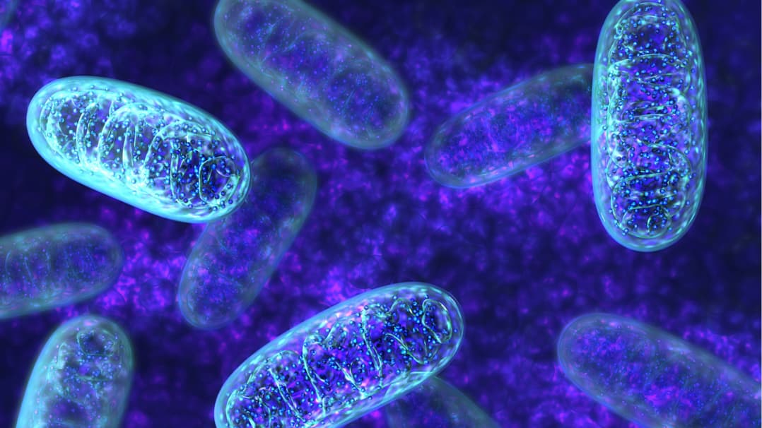
NAD+ is a core molecule of cellular function. NAD+ has a central role in the production of cellular energy as adenosine triphosphate (ATP), and therefore, in powering all cellular functions that rely on ATP. NAD+ is also essential for redox homeostasis and cell signaling pathways involved in maintaining cellular health [1]. NAD+ levels decrease with aging and this decline is associated with many dysfunctions that develop as we age. Interventions aimed at maintaining NAD+ levels have the potential to support the processes that rely on NAD+ and consequently help to prevent or mitigate age-related dysfunctions and promote healthy aging.
Nicotinamide mononucleotide (NMN) and nicotinamide riboside (NR) are two NAD+ biosynthesis precursors that can enhance NAD+ levels. Boosting NAD+ levels through NMN and/or NR has been proposed as a way to potentially mitigate the physiological decline in NAD+ levels associated with aging and to help prevent or alleviate age-related dysfunctions.
A question often asked is “Which is better: NMN or NR?” It may not be possible to provide a definite answer at the moment, but it’s worth understanding these molecules and exploring the similarities and differences between NMN and NR. But to understand NMN vs NR, we must first understand NAD+.
Understanding NAD+
NAD+ is the oxidized form of nicotinamide adenine dinucleotide (NAD), a coenzyme form of vitamin B3, meaning it supports the activity of enzymes. The NAD molecule consists of nicotinamide mononucleotide (NMN) bound to adenosine monophosphate (AMP). Vitamin B3 is also known as niacin, which is one of its forms (also known as nicotinic acid). Niacinamide (or nicotinamide) is another form of Vitamin B3.
NAD+ and its reduced form NADH have key roles in redox homeostasis. They are converted back and forth between these two forms in redox reactions (i.e., reactions where electron transfer occurs). These molecules must be maintained at a high NAD+/NADH ratio (i.e., higher levels of NAD+ than NADH) to maintain healthy mitochondrial and cellular function [2,3].

Figure 1. NAD+ ←→ NADH Redox Reaction
One of the main roles of NAD+ is its involvement in cellular energy pathways that produce ATP. Glycolysis (which breaks down glucose), beta-oxidation (which breaks down fatty acids), and the citric acid cycle (or Krebs cycle, which extracts electrons from carbon units derived from other pathways) produce a small amount of ATP at the cost of converting NAD+ molecules to NADH, which acts as an electron carrier. In oxidative phosphorylation, NADH feeds electrons into the electron transport chain to power the production of great amounts of ATP, being converted back to NAD+ in the process. This regeneration of NAD+ helps cells to maintain a high NAD+/NADH ratio.
NAD+ also participates in signaling pathways and processes essential for cell health [4]. NAD+ is a co-substrate of NAD-dependent enzymes such as PARPs and sirtuins that mediate important processes such as protection of mitochondrial function, metabolic adjustments, redox homeostasis, DNA repair and genomic stability, apoptosis, cell differentiation, and survival, among others [5–8].
How to Boost NAD+
These NAD+-dependent reactions consume NAD+ and produce niacinamide as a byproduct [9]. To maintain a high NAD+/NADH ratio, niacinamide is recycled back to NAD+ through one of the pathways of NAD+ production, known as the salvage pathway.
NAD+ can also be produced from tryptophan (de novo synthesis pathway) and niacin (Preiss-Handler pathway). So NAD+ is continuously synthesized, metabolized, and recycled in the cell through different pathways. Sustaining stable levels of NAD+ is important because, as a result of its actions, NAD+ plays a key role in maintaining healthy cells and tissues. Maintaining adequate levels of NAD+ is therefore crucial for maintaining our overall health.
However, NAD+ levels in tissues decline as we age [10–12]. This decline is believed to contribute to age-related physiological decline. The decline is one of the primary reasons why there has been so much scientific interest in compounds like NMN and NR for boosting NAD+.
The decline in NAD+ levels in tissues as we age is one of the primary reasons there has been so much scientific interest in compounds like NMN and NR for boosting NAD.
Studies with aged animals have shown that restoration of NAD+ levels mitigates many age-related changes by enhancing mitochondrial function and energy metabolism, restoring cellular defenses against oxidative stress, promoting DNA repair, and modulating cellular senescence. By doing so, restoring NAD+ can ameliorate systemic and tissue dysfunction and promote healthspan [13,14].
These outcomes resulted in a big increase in interest in strategies to increase NAD+ levels, with the hope being that boosting NAD+ levels in humans might have similar functional benefits. One way to do so is by getting more NAD+ precursors in the diet to support NAD+ production. Niacin and niacinamide are the most commonly used forms to do so. NMN and NR are two new promising possibilities: they are both precursors for NAD+ biosynthesis that are capable of boosting NAD+ production.
NMN (Nicotinamide Mononucleotide)
NMN consists of a niacinamide molecule added to a ribose sugar and a phosphate group. There is still limited data on NMN content in foods, but one study has determined that NMN is found in small amounts in fruits and vegetables such as edamame, avocado, broccoli, cucumber, cabbage, and tomato, and in smaller amounts in meat and shrimp [15].

Figure 2. NMN structure
NMN is a precursor for NAD+ biosynthesis through the salvage pathway. As mentioned above, the salvage pathway recycles niacinamide (NAM) which is produced as a byproduct of the consumption of NAD+ in NAD+-dependent signaling reactions. NMN is an intermediate in the pathway that turns NAM into NAD+ [16].
Preclinical studies have demonstrated that oral NMN supplementation enhances NAD+ levels in several peripheral tissues, including the liver, kidney, white adipose tissue, pancreas, lung, heart, and skeletal muscle [9,16–20]. NMN mitigated oxidative stress, and DNA damage, and supported healthy insulin secretion, mitochondrial function, energy metabolism, metabolic health, immune responses, and physical activity. NMN mitigated cardiovascular dysfunctions and skeletal muscle aging and supported neuroprotective functions (protected from cognitive impairment, neuronal death, and synaptic loss) [15,16,21–28].
NMN Studies
To date, studies in humans are still limited, but NMN supplementation has been shown to enhance levels of NAD+ in the blood [29–34]. Some physiological benefits have been observed in clinical trials. NMN supported muscle insulin sensitivity and remodeling in overweight women [29], enhanced physical performance in healthy middle-aged and older adults [30,34], and supported healthy blood cholesterol levels, body weight, and diastolic blood pressure in overweight adults over 45 years [32]. On the other hand, NMN had no significant effects on arterial health, insulin sensitivity, body composition, cognitive testing, cardiometabolic markers, sleep quality, fatigue, and physical performance in healthy middle-aged and older individuals [32,34–38].
NMN supplementation has been shown to enhance levels of NAD+ in the blood.
In clinical trials, NMN did not cause any serious side effects, any differences in adverse events compared to placebo, nor any meaningful differences in clinical analyses; NMN was well tolerated in doses up to 1250 mg for up to 4 weeks [30,31,39] and at lower doses of 250 -300 mg/day for up to 24 weeks [33,37,40].
NR (Nicotinamide Riboside)
NR consists of a niacinamide molecule added to a ribose sugar. Data on NR content in foods is very limited, but it has been found in small amounts in cow milk [41] and Brewer's yeast [42].

Figure 3. NR structure
Like NMN, NR is a precursor for NAD+ biosynthesis through the salvage pathway. But unlike NMN, it is not an intermediate of the pathway. NR enters the salvage pathway by being metabolized to NMN or niacinamide, which can then yield NAD+ [16].
NR Studies
Preclinical studies have shown that oral NR supplementation enhances levels of NAD+ in the liver, muscle, and brown adipose tissue [43,44]. NR demonstrated protective effects on cognitive function, hippocampal neurons, and synaptic plasticity, upregulated BDNF, promoted neurogenesis, and modulated microglial activation. NR also supported oxidative metabolism, mitochondrial content, glucose tolerance, insulin sensitivity, liver structure, and cardiac function [44–49].
Studies in humans with NR are also still limited, but NR supplementation has been shown to enhance levels of NAD+ in the blood [50–54]. A clinical study with NR as NIAGEN® showed an enhancement in blood NAD+ levels within two weeks that was maintained throughout the eight weeks of the study. The NAD+ enhancement was dose-dependent (22%, 51%, and 142% with 100, 300, and 1000 mg, respectively) [53].
NMN vs. NR: A Comparative Analysis
NMN and NR are related molecules with several things in common but with important differences.
NMN and NR are structurally identical except for an additional phosphate group in the structure of NMN. This phosphate group makes the NMN molecule larger, resulting in NMN likely having to be converted into NR to cross cell membranes. On the other hand, although NMN and NR are both precursors for NAD+ biosynthesis through the salvage pathway, NR must be converted into NMN to flow through the salvage pathway to produce NAD+.
There are also differences in how NMN and NR are absorbed in the gut. Although the mechanisms are not yet fully elucidated, data from animal studies has revealed a few possible processes of NMN and NR absorption and NAD+ enhancement.
Oral NMN may be absorbed and enhance NAD+ levels in different ways: 1) NMN may be absorbed directly, as suggested by the identification of a possible NMN transporter in the gut [66] (although these findings have been challenged [67,68]), and then be metabolized to NAD+ through the salvage pathway in the liver; 2) NMN may be dephosphorylated to NR before absorption, which may then cross cell membranes through transporters [17,69] and be reconverted to NMN to be metabolized to NAD+ by the salvage pathway in the liver); 3) NMN may be partially deamidated by the gut microbiota to nicotinic acid mononucleotide (NAMN), which may then be absorbed and metabolized to NAD+ through the Preiss-Handler pathway in the liver [70].
Oral NR may also be absorbed and enhance NAD+ levels in different ways: 1) NR may be directly absorbed from the gut through cell membrane transporters and contribute to NAD+ generation in the liver through the salvage pathway via conversion back to NMN [71]; 2) NR may be metabolized by the gut microbiota by being deaminated into nicotinic acid riboside (NAR) or hydrolyzed to nicotinamide and then deaminated to niacin; both NAR and niacin can then contribute to the generation of NAD+ through the Preiss–Handler pathway in the liver [72,73].
How each of these processes contributes to NMN and NR absorption and if they’re similar in humans remains to be determined. But regardless of the process, orally administered NR and NMN seem to be nearly fully metabolized to NAD+ and niacinamide in the gut and the liver following absorption [15,65,74]. This means that their impact on NAD+ levels in other tissues is the result of their conversion into niacinamide, the primary circulating form of vitamin B3, which is then distributed to other tissues through the blood and metabolized to NAD+ [15,65,74].
Choosing Between NMN and NR
The absence of studies that directly compare NR vs NMN makes it hard to determine which is better. Both can boost NAD+ in humans, but both still need more research to determine the extent of their benefits, the optimal dose, and in which contexts one may be better than the other. Would taking both together produce benefits beyond what taking one alone does? The answer to that question is also unknown.
NAD+ can be produced through different pathways and the relevance of each pathway may vary based on the biosynthetic capacity of enzymes in different tissues. This capacity can also change with age or with health status [79].
With Qualia formulations, we believe a better approach for long-term health is to support the functional redundancy inherent in the human body for NAD+ maintenance. This entails providing several substrates for NAD+ biosynthesis, as well as supporting rate-limiting steps in the different pathways. This has been our approach in designing Qualia NAD+. You can learn more about it in The Formulator's View of the Qualia NAD+ Ingredients.
However, at the moment, the FDA has determined that NMN is excluded from the definition of dietary NAD+ supplementation because they had previously authorized it for investigation as a new drug. This is the reason why NMN is not in Qualia NAD+. But we did include three precursor forms of Vitamin B3: NR as NIAGEN®, niacin, and niacinamide.

*These statements have not been evaluated by the Food and Drug Administration. This product is not intended to diagnose, treat, cure, or prevent any disease.
References
[1]A.J. Covarrubias, R. Perrone, A. Grozio, E. Verdin, Nat. Rev. Mol. Cell Biol. 22 (2021) 119–141.
[2]M.E. Tischler, D. Friedrichs, K. Coll, J.R. Williamson, Arch. Biochem. Biophys. 184 (1977) 222–236.
[3]D.H. Williamson, P. Lund, H.A. Krebs, Biochem. J 103 (1967) 514–527.
[4]A.R. Mendelsohn, J.W. Larrick, Rejuvenation Res. 20 (2017) 244–247.
[5]M. Pittelli, R. Felici, V. Pitozzi, L. Giovannelli, E. Bigagli, F. Cialdai, G. Romano, F. Moroni, A. Chiarugi, Mol. Pharmacol. 80 (2011) 1136–1146.
[6]Z. Herceg, Z.Q. Wang, Mutat. Res. 477 (2001) 97–110.
[7]H. Zhang, D. Ryu, Y. Wu, K. Gariani, X. Wang, P. Luan, D. D’Amico, E.R. Ropelle, M.P. Lutolf, R. Aebersold, K. Schoonjans, K.J. Menzies, J. Auwerx, Science 352 (2016) 1436–1443.
[8]R.H. Houtkooper, E. Pirinen, J. Auwerx, Nat. Rev. Mol. Cell Biol. 13 (2012) 225–238.
[9]A.P. Gomes, N.L. Price, A.J.Y. Ling, J.J. Moslehi, M.K. Montgomery, L. Rajman, J.P. White, J.S. Teodoro, C.D. Wrann, B.P. Hubbard, E.M. Mercken, C.M. Palmeira, R. de Cabo, A.P.
Rolo, N. Turner, E.L. Bell, D.A. Sinclair, Cell 155 (2013) 1624–1638.
[10]J. Clement, M. Wong, A. Poljak, P. Sachdev, N. Braidy, Rejuvenation Res. (2018).
[11]S.-I. Imai, L. Guarente, Trends Cell Biol. 24 (2014) 464–471.
[12]E. Verdin, Science 350 (2015) 1208–1213.
[13]C.F. Lee, A. Caudal, L. Abell, G.A. Nagana Gowda, R. Tian, Sci. Rep. 9 (2019) 3073.
[14]E.F. Fang, S. Lautrup, Y. Hou, T.G. Demarest, D.L. Croteau, M.P. Mattson, V.A. Bohr, Trends Mol. Med. 23 (2017) 899–916.
[15]K.F. Mills, S. Yoshida, L.R. Stein, A. Grozio, S. Kubota, Y. Sasaki, P. Redpath, M.E. Migaud, R.S. Apte, K. Uchida, J. Yoshino, S.-I. Imai, Cell Metab. 24 (2016) 795–806.
[16]J. Yoshino, J.A. Baur, S.-I. Imai, Cell Metab. 27 (2018) 513–528.
[17]A.A. Sauve, Q. Wang, N. Zhang, S. Kang, A. Rathmann, Y. Yang, Int. J. Mol. Sci. 24 (2023).
[18]J. Yoshino, K.F. Mills, M.J. Yoon, S.-I. Imai, Cell Metab. 14 (2011) 528–536.
[19]T. Yamamoto, J. Byun, P. Zhai, Y. Ikeda, S. Oka, J. Sadoshima, PLoS One 9 (2014) e98972.
[20]A.S. Martin, D.M. Abraham, K.A. Hershberger, D.P. Bhatt, L. Mao, H. Cui, J. Liu, X. Liu, M.J. Muehlbauer, P.A. Grimsrud, J.W. Locasale, R.M. Payne, M.D. Hirschey, JCI Insight 2 (2017).
[21]J. Yoshino, K.F. Mills, M.J. Yoon, S.-I. Imai, Cell Metab. 14 (2011) 528–536.
[22]P.W. Caton, J. Kieswich, M.M. Yaqoob, M.J. Holness, M.C. Sugden, Diabetologia 54 (2011) 3083–3092.
[23]X. Wang, X. Hu, Y. Yang, T. Takata, T. Sakurai, Brain Res. 1643 (2016) 1–9.
[24]Z. Yao, W. Yang, Z. Gao, P. Jia, Neurosci. Lett. 647 (2017) 133–140.
[25]N.E. de Picciotto, L.B. Gano, L.C. Johnson, C.R. Martens, A.L. Sindler, K.F. Mills, S.-I. Imai, D.R. Seals, Aging Cell 15 (2016) 522–530.
[26]Ł. Mateuszuk, R. Campagna, B. Kutryb-Zając, K. Kuś, E.M. Słominska, R.T. Smolenski, S. Chlopicki, Biochem. Pharmacol. 178 (2020) 114019.
[27]R. Zhang, Y. Shen, L. Zhou, P. Sangwung, H. Fujioka, L. Zhang, X. Liao, J. Mol. Cell. Cardiol. 112 (2017) 64–73.
[28]A.R. Mendelsohn, J.W. Larrick, Rejuvenation Res. 17 (2014) 62–69.
[29]M. Yoshino, J. Yoshino, B.D. Kayser, G.J. Patti, M.P. Franczyk, K.F. Mills, M. Sindelar, T. Pietka, B.W. Patterson, S.-I. Imai, S. Klein, Science 372 (2021) 1224–1229.
[30]L. Yi, A.B. Maier, R. Tao, Z. Lin, A. Vaidya, S. Pendse, S. Thasma, N. Andhalkar, G. Avhad, V. Kumbhar, Geroscience 45 (2023) 29–43.
[31]K.M. Pencina, S. Lavu, M. Dos Santos, Y.M. Beleva, M. Cheng, D. Livingston, S. Bhasin, J. Gerontol. A Biol. Sci. Med. Sci. 78 (2023) 90–96.
[32]K.M. Pencina, R. Valderrabano, B. Wipper, A.R. Orkaby, K.F. Reid, T. Storer, A.P. Lin, S. Merugumala, L. Wilson, N. Latham, C. Ghattas-Puylara, N.E. Ozimek, M. Cheng, A. Bhargava, Y. Memish-Beleva, B. Lawney, S. Lavu, P.M. Swain, R.S. Apte, D.A. Sinclair, D. Livingston, S. Bhasin, J. Clin. Endocrinol. Metab. 108 (2023) 1968–1980.
[33]K. Okabe, K. Yaku, Y. Uchida, Y. Fukamizu, T. Sato, T. Sakurai, K. Tobe, T. Nakagawa, Front Nutr 9 (2022) 868640.
[34]M. Igarashi, Y. Nakagawa-Nagahama, M. Miura, K. Kashiwabara, K. Yaku, M. Sawada, R. Sekine, Y. Fukamizu, T. Sato, T. Sakurai, J. Sato, K. Ino, N. Kubota, T. Nakagawa, T. Kadowaki, T. Yamauchi, NPJ Aging 8 (2022) 5.
[35]T. Katayoshi, S. Uehata, N. Nakashima, T. Nakajo, N. Kitajima, M. Kageyama, K. Tsuji-Naito, Sci. Rep. 13 (2023) 2786.
[36]L. Yi, A.B. Maier, R. Tao, Z. Lin, A. Vaidya, S. Pendse, S. Thasma, N. Andhalkar, G. Avhad, V. Kumbhar, Geroscience 45 (2023) 29–43.
[37]H. Huang, Front Aging 3 (2022) 851698.
[38]M. Kim, J. Seol, T. Sato, Y. Fukamizu, T. Sakurai, T. Okura, Nutrients 14 (2022).
[39]Y. Fukamizu, Y. Uchida, A. Shigekawa, T. Sato, H. Kosaka, T. Sakurai, Sci. Rep. 12 (2022) 14442.
[40]H. Akasaka, H. Nakagami, K. Sugimoto, Y. Yasunobe, T. Minami, T. Fujimoto, K. Yamamoto, C. Hara, A. Shiraki, K. Nishida, K. Asano, M. Kanou, K. Yamana, S.-I. Imai, H. Rakugi, Geriatr. Gerontol. Int. 23 (2023) 38–43.
[41]S.A. Trammell, L. Yu, P. Redpath, M.E. Migaud, C. Brenner, J. Nutr. 146 (2016) 957–963.
[42]E.S. Holdsworth, D.V. Kaufman, E. Neville, Br. J. Nutr. 65 (1991) 285–299.
[43]S.A.J. Trammell, M.S. Schmidt, B.J. Weidemann, P. Redpath, F. Jaksch, R.W. Dellinger, Z. Li, E.D. Abel, M.E. Migaud, C. Brenner, Nat. Commun. 7 (2016) 12948.
[44]C. Cantó, R.H. Houtkooper, E. Pirinen, D.Y. Youn, M.H. Oosterveer, Y. Cen, P.J. Fernandez-Marcos, H. Yamamoto, P.A. Andreux, P. Cettour-Rose, K. Gademann, C. Rinsch, K. Schoonjans, A.A. Sauve, J. Auwerx, Cell Metab. 15 (2012) 838–847.
[45]Y. Hou, S. Lautrup, S. Cordonnier, Y. Wang, D.L. Croteau, E. Zavala, Y. Zhang, K. Moritoh, J.F. O’Connell, B.A. Baptiste, T.V. Stevnsner, M.P. Mattson, V.A. Bohr, Proc. Natl. Acad. Sci. U. S. A. 115 (2018) E1876–E1885.
[46]B. Gong, Y. Pan, P. Vempati, W. Zhao, L. Knable, L. Ho, J. Wang, M. Sastre, K. Ono, A.A. Sauve, G.M. Pasinetti, Neurobiol. Aging 34 (2013) 1581–1588.
[47]Y. Jiang, Y. Liu, M. Gao, M. Xue, Z. Wang, H. Liang, Food Funct. 11 (2020) 378–391.
[48]K. Gariani, K.J. Menzies, D. Ryu, C.J. Wegner, X. Wang, E.R. Ropelle, N. Moullan, H. Zhang, A. Perino, V. Lemos, B. Kim, Y.-K. Park, A. Piersigilli, T.X. Pham, Y. Yang, C.S. Ku, S.I. Koo, A. Fomitchova, C. Cantó, K. Schoonjans, A.A. Sauve, J.-Y. Lee, J. Auwerx, Hepatology 63 (2016) 1190–1204.
[49]N. Diguet, S.A.J. Trammell, C. Tannous, R. Deloux, J. Piquereau, N. Mougenot, A. Gouge, M. Gressette, B. Manoury, J. Blanc, M. Breton, J.-F. Decaux, G.G. Lavery, I. Baczkó, J. Zoll, A. Garnier, Z. Li, C. Brenner, M. Mericskay, Circulation 137 (2018) 2256–2273.
[50]S.E. Airhart, L.M. Shireman, L.J. Risler, G.D. Anderson, G.A. Nagana Gowda, D. Raftery, R. Tian, D.D. Shen, K.D. O’Brien, PLoS One 12 (2017) e0186459.
[51]C.R. Martens, B.A. Denman, M.R. Mazzo, M.L. Armstrong, N. Reisdorph, M.B. McQueen, M. Chonchol, D.R. Seals, Nat. Commun. 9 (2018) 1286.
[52]Y.S. Elhassan, K. Kluckova, R.S. Fletcher, M.S. Schmidt, A. Garten, C.L. Doig, D.M. Cartwright, L. Oakey, C.V. Burley, N. Jenkinson, M. Wilson, S.J.E. Lucas, I. Akerman, A. Seabright, Y.-C. Lai, D.A. Tennant, P. Nightingale, G.A. Wallis, K.N. Manolopoulos, C. Brenner, A. Philp, G.G. Lavery, Cell Rep. 28 (2019) 1717–1728.e6.
[53]D. Conze, C. Brenner, C.L. Kruger, Sci. Rep. 9 (2019) 9772.
[54]H.A.K. Lapatto, M. Kuusela, A. Heikkinen, M. Muniandy, B.W. van der Kolk, S. Gopalakrishnan, N. Pöllänen, M. Sandvik, M.S. Schmidt, S. Heinonen, S. Saari, J. Kuula, A. Hakkarainen, J. Tampio, T. Saarinen, M.-R. Taskinen, N. Lundbom, P.-H. Groop, M. Tiirola, P. Katajisto, M. Lehtonen, C. Brenner, J. Kaprio, S. Pekkala, M. Ollikainen, K.H. Pietiläinen, E. Pirinen, Sci Adv 9 (2023) eadd5163.
[55]B. Brakedal, C. Dölle, F. Riemer, Y. Ma, G.S. Nido, G.O. Skeie, A.R. Craven, T. Schwarzlmüller, N. Brekke, J. Diab, L. Sverkeli, V. Skjeie, K. Varhaug, O.-B. Tysnes, S. Peng, K. Haugarvoll, M. Ziegler, R. Grüner, D. Eidelberg, C. Tzoulis, Cell Metab. 34 (2022) 396–407.e6.
[56]A. Ahmadi, G. Begue, A.P. Valencia, J.E. Norman, B. Lidgard, B.J. Bennett, M.P. Van Doren, D.J. Marcinek, S. Fan, D.K. Prince, J. Gamboa, J. Himmelfarb, I.H. de Boer, B.R. Kestenbaum, B. Roshanravan, JCI Insight 8 (2023).
[57]C.F. Dolopikou, I.A. Kourtzidis, N.V. Margaritelis, I.S. Vrabas, I. Koidou, A. Kyparos, A.A. Theodorou, V. Paschalis, M.G. Nikolaidis, Eur. J. Nutr. 59 (2020) 505–515.
[58]O.L. Dollerup, S. Chubanava, M. Agerholm, S.D. Søndergård, A. Altıntaş, A.B. Møller, K.F. Høyer, S. Ringgaard, H. Stødkilde-Jørgensen, G.G. Lavery, R. Barrès, S. Larsen, C. Prats, N. Jessen, J.T. Treebak, J. Physiol. 598 (2020) 731–754.
[59]C.M.E. Remie, K.H.M. Roumans, M.P.B. Moonen, N.J. Connell, B. Havekes, J. Mevenkamp, L. Lindeboom, V.H.W. de Wit, T. van de Weijer, S.A.B.M. Aarts, E. Lutgens, B.V. Schomakers, H.L. Elfrink, R. Zapata-Pérez, R.H. Houtkooper, J. Auwerx, J. Hoeks, V.B. Schrauwen-Hinderling, E. Phielix, P. Schrauwen, Am. J. Clin. Nutr. 112 (2020) 413–426.
[60]O.L. Dollerup, S.A.J. Trammell, B. Hartmann, J.J. Holst, B. Christensen, N. Møller, M.P. Gillum, J.T. Treebak, N. Jessen, J. Clin. Endocrinol. Metab. 104 (2019) 5703–5714.
[61]O.L. Dollerup, B. Christensen, M. Svart, M.S. Schmidt, K. Sulek, S. Ringgaard, H. Stødkilde-Jørgensen, N. Møller, C. Brenner, J.T. Treebak, N. Jessen, Am. J. Clin. Nutr. 108 (2018) 343–353.[62]B. Stocks, S.P. Ashcroft, S. Joanisse, L.C. Dansereau, Y.C. Koay, Y.S. Elhassan, G.G. Lavery, L.-E. Quek, J.F. O’Sullivan, A.M. Philp, G.A. Wallis, A. Philp, J. Physiol. 599 (2021) 1513–1531.
[63]A.A. Peluso, A.T. Lundgaard, P. Babaei, F. Mousovich-Neto, A.L. Rocha, M.V. Damgaard, E.G. Bak, T. Gnanasekaran, O.L. Dollerup, S.A.J. Trammell, T.S. Nielsen, T. Kern, C.B. Abild, K. Sulek, T. Ma, Z. Gerhart-Hines, M.P. Gillum, M. Arumugam, C. Ørskov, D. McCloskey, N. Jessen, M.J. Herrgård, M.A.S. Mori, J.T. Treebak, NPJ Aging 9 (2023) 7.
[64]C.R. Martens, B.A. Denman, M.R. Mazzo, M.L. Armstrong, N. Reisdorph, M.B. McQueen, M. Chonchol, D.R. Seals, Nat. Commun. 9 (2018) 1286.
[65]S.A.J. Trammell, M.S. Schmidt, B.J. Weidemann, P. Redpath, F. Jaksch, R.W. Dellinger, Z. Li, E.D. Abel, M.E. Migaud, C. Brenner, Nat. Commun. 7 (2016) 12948.
[66]A. Grozio, K.F. Mills, J. Yoshino, S. Bruzzone, G. Sociali, K. Tokizane, H.C. Lei, R. Cunningham, Y. Sasaki, M.E. Migaud, S.-I. Imai, Nat Metab 1 (2019) 47–57.
[67]M.S. Schmidt, C. Brenner, Nat Metab 1 (2019) 660–661.
[68]A. Grozio, K. Mills, J. Yoshino, S. Bruzzone, G. Sociali, K. Tokizane, H.C. Lei, Y. Sasaki, M. Migaud, S.-I. Imai, Nat Metab 1 (2019) 662–665.
[69]J. Ratajczak, M. Joffraud, S.A.J. Trammell, R. Ras, N. Canela, M. Boutant, S.S. Kulkarni, M. Rodrigues, P. Redpath, M.E. Migaud, J. Auwerx, O. Yanes, C. Brenner, C. Cantó, Nat. Commun. 7 (2016) 13103.
[70]L. Galeazzi, P. Bocci, A. Amici, L. Brunetti, S. Ruggieri, M. Romine, S. Reed, A.L. Osterman, D.A. Rodionov, L. Sorci, N. Raffaelli, J. Biol. Chem. 286 (2011) 40365–40375.
[71]M. Mehmel, N. Jovanović, U. Spitz, Nutrients 12 (2020).
[72]K. Yaku, S. Palikhe, H. Izumi, T. Yoshida, K. Hikosaka, F. Hayat, M. Karim, T. Iqbal, Y. Nitta, A. Sato, M.E. Migaud, K. Ishihara, H. Mori, T. Nakagawa, Nat. Commun. 12 (2021) 6767.
[73]I. Shats, J.G. Williams, J. Liu, M.V. Makarov, X. Wu, F.B. Lih, L.J. Deterding, C. Lim, X. Xu, T.A. Randall, E. Lee, W. Li, W. Fan, J.-L. Li, M. Sokolsky, A.V. Kabanov, L. Li, M.E. Migaud, J.W. Locasale, X. Li, Cell Metab. 31 (2020) 564–579.e7.
[74]L. Liu, X. Su, W.J. Quinn 3rd, S. Hui, K. Krukenberg, D.W. Frederick, P. Redpath, L. Zhan, K. Chellappa, E. White, M. Migaud, T.J. Mitchison, J.A. Baur, J.D. Rabinowitz, Cell Metab. 27 (2018) 1067–1080.e5.
[75]E.F. Fang, H. Kassahun, D.L. Croteau, M. Scheibye-Knudsen, K. Marosi, H. Lu, R.A. Shamanna, S. Kalyanasundaram, R.C. Bollineni, M.A. Wilson, W.B. Iser, B.N. Wollman, M. Morevati, J. Li, J.S. Kerr, Q. Lu, T.B. Waltz, J. Tian, D.A. Sinclair, M.P. Mattson, H. Nilsen, V.A. Bohr, Cell Metab. 24 (2016) 566–581.
[76]E.F. Fang, M. Scheibye-Knudsen, L.E. Brace, H. Kassahun, T. SenGupta, H. Nilsen, J.R. Mitchell, D.L. Croteau, V.A. Bohr, Cell 157 (2014) 882–896.
[77]E.F. Fang, Y. Hou, S. Lautrup, M.B. Jensen, B. Yang, T. SenGupta, D. Caponio, R. Khezri, T.G. Demarest, Y. Aman, D. Figueroa, M. Morevati, H.-J. Lee, H. Kato, H. Kassahun, J.-H. Lee, D. Filippelli, M.N. Okur, A. Mangerich, D.L. Croteau, Y. Maezawa, C.A. Lyssiotis, J. Tao, K. Yokote, T.E. Rusten, M.P. Mattson, H. Jasper, H. Nilsen, V.A. Bohr, Nat. Commun. 10 (2019) 5284.
[78]M.N. Okur, E.F. Fang, E.M. Fivenson, V. Tiwari, D.L. Croteau, V.A. Bohr, Aging Cell 19 (2020) e13268.
[79]K.L. Bogan, C. Brenner, Annu. Rev. Nutr. 28 (2008) 115–130.